
In short: the three modes show broadly the same things, but not at the same depth. Immersion (non-polarized light) excels at the surface; polarized light sees deeper and gives its best images in contact mode. The two are complementary.
When choosing a dermatoscope, the question always comes up: polarized or non-polarized? And where does the immersion fluid come in? This article explains the principle in simple terms, what each mode shows best, and what the studies say. It is intended for dermatologists as much as for general practitioners or podiatrists starting out in dermoscopy.
The starting problem: the skin acts like a mirror
Every dermatoscope has to solve the same problem: glare. When light hits the skin, part of it bounces straight off the stratum corneum. This bright white reflection hides what lies beneath, exactly like the sun on a window.
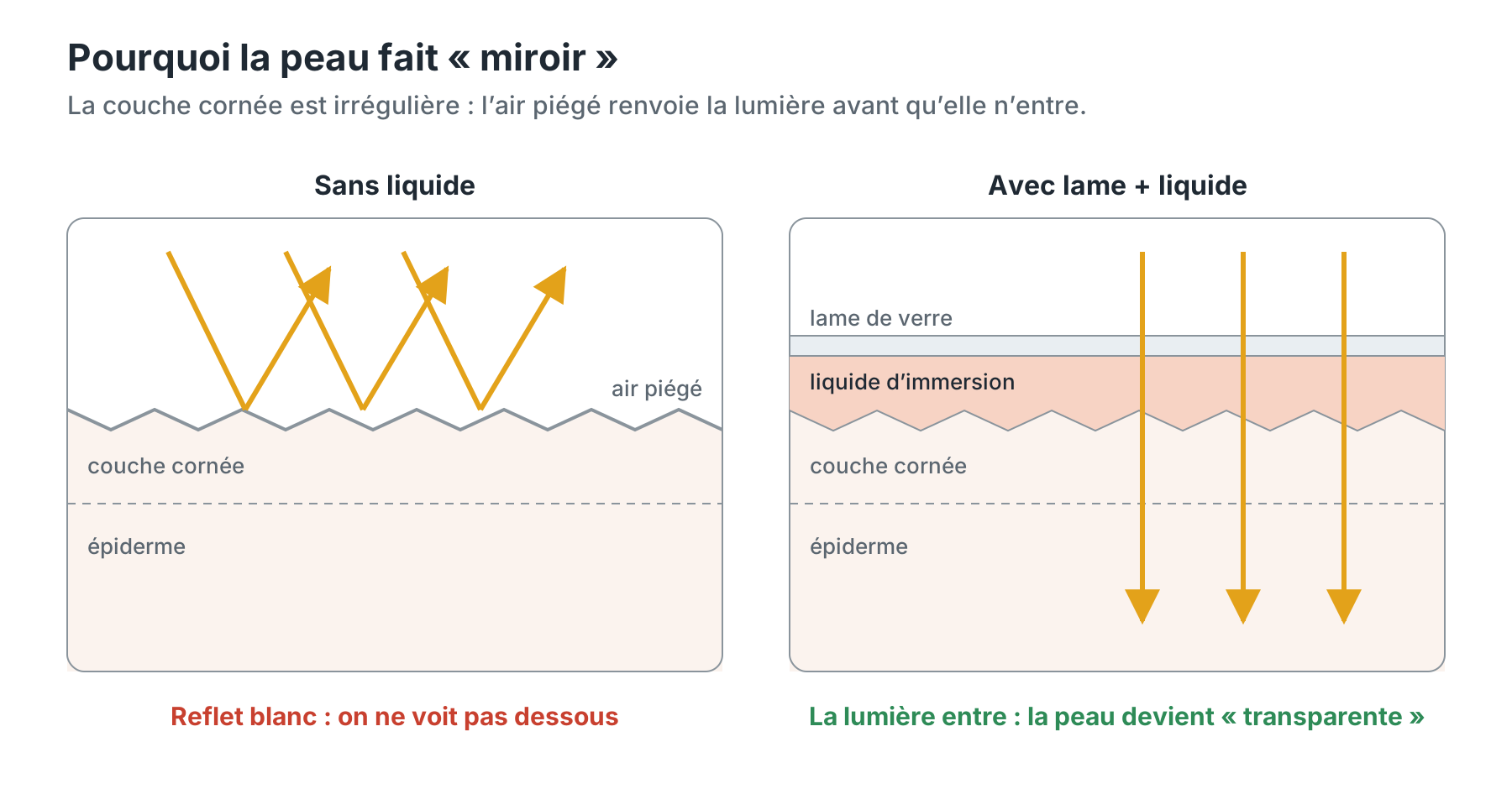
To see the pigments and vessels below the surface, this glare must therefore be eliminated. There are two ways to do this:
- remove the glare at the source: a glass plate and a fluid are placed on the skin so that light enters instead of bouncing off. This is non-polarized immersion dermoscopy;
- filter the glare on the way back: two polarizing filters block the light reflected at the surface. This is polarized dermoscopy, with or without contact.
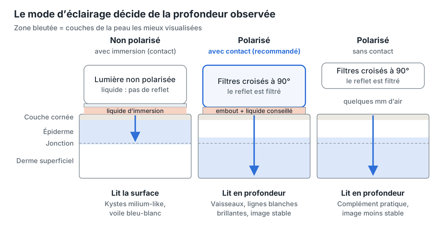
There are therefore three modes: non-polarized contact (immersion), polarized contact, and polarized non-contact. This is the classification used by the clinical reference DermNet.
Non-polarized immersion dermoscopy
This is the original mode: excellent for the skin surface. The illuminating light goes in all directions. To keep it from bouncing back, an interface fluid (ultrasound gel, oil, alcohol or water) is applied and the contact plate is placed on the lesion. The fluid fills in the irregularities of the stratum corneum and drives out the air: the skin becomes “transparent”.
What this mode shows best: the superficial epidermis down to the dermoepidermal junction. Milia-like cysts and comedo-like openings of seborrheic keratoses are very clear, as is the blue-white veil.
Its day-to-day constraints:
- it requires fluid and direct contact, for every lesion;
- the contact plate must be disinfected between patients;
- the pressure of the plate blanches small vessels: vascular structures and pink color are less visible;
- air bubbles trapped under the plate interfere with reading.
Polarized dermoscopy
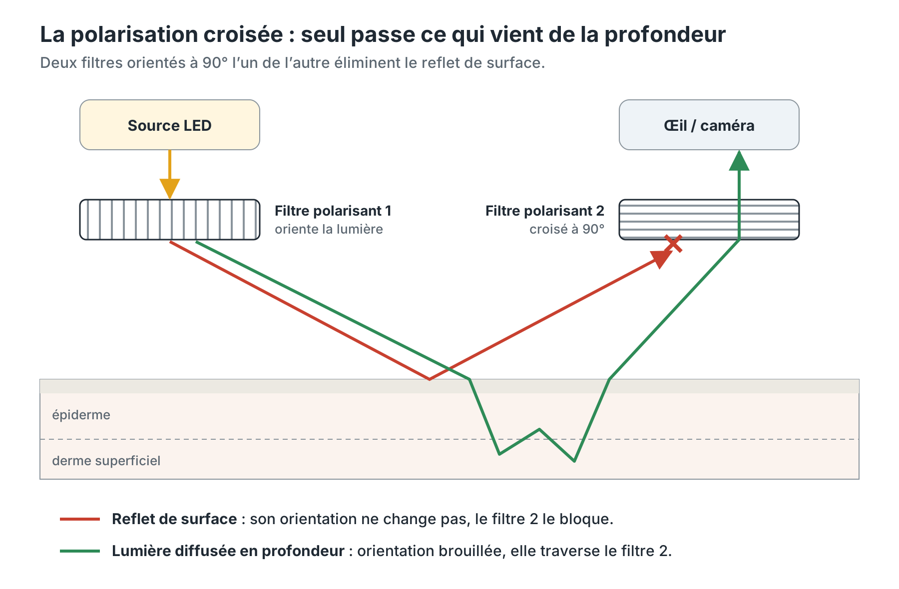
It sees deeper, and is preferably used in contact. The light passes through a first filter that orients it in a single plane. A second filter, placed at 90° in front of the lens, blocks all the light that bounces off the surface without changing orientation. Only light that has penetrated the skin and been scattered there gets through. This is called cross-polarization.
Result: the glare disappears, and the image comes mainly from the dermoepidermal junction and the superficial dermis.
Two ways to use it:
- with contact (recommended): the tip is placed on the skin, ideally with a drop of fluid. The device is stable, focus is constant and the image is clearly better. This is the mode to favor day to day;
- without contact: the device is held a few millimeters from the skin. No pressure on the vessels and no tip to disinfect, but the image is more sensitive to movement and stray reflections. Best kept for areas where contact is difficult or for observing a fragile vascular lesion.
Polarized contact with fluid: the best compromise. Adding an immersion fluid in polarized mode does not cancel out the advantages of polarization. According to the International Dermoscopy Society, a fluid such as 70% alcohol can even improve image quality, probably because it lets more light through the stratum corneum (dermoscopedia). It is also what allows a hybrid device to be used in both modes without lifting it from the skin.
What this mode shows best: vessels and pink color, as well as shiny white structures (chrysalis lines, rosettes). The latter, related to dermal collagen, are visible only in polarized light. They are important clues to melanoma and basal cell carcinoma.
What each mode shows best
Remember a simple rule: surface = non-polarized, depth = polarized. The table below reproduces the grid published by the International Dermoscopy Society on dermoscopedia.
| Color or structure | Non-polarized (immersion) | Polarized |
|---|---|---|
| Blood vessels |
| |
| Pink / red color |
|
|
| Shiny white structures (chrysalis) | / |
|
| Melanin (brown pigment) |
|
|
| Milia-like cysts |
| / |
| Blue-white veil |
|
|
| Granularity (peppering) |
|
|
In practice, the three modes also differ in day-to-day use:
| Criterion | Non-polarized with immersion | Polarized contact (recommended) | Polarized non-contact |
|---|---|---|---|
| Interface fluid | Required | Recommended | None |
| Contact with the skin | Yes | Yes | No |
| Image stability and sharpness | Very good | Very good | More sensitive to movement |
| Pressure on vessels | Yes | Slight | None |
| Disinfection between patients | Contact plate | Tip | No contact |
| Depth observed | Epidermis → junction | Junction → superficial dermis | Junction → superficial dermis |
The “blink sign”. On a hybrid device, switching from one mode to the other makes certain structures “blink”: they appear in one mode and disappear in the other. This contrast helps identify them. Note: to benefit from it, the device must be used in contact and with a fluid; otherwise only polarized mode gives a usable image.
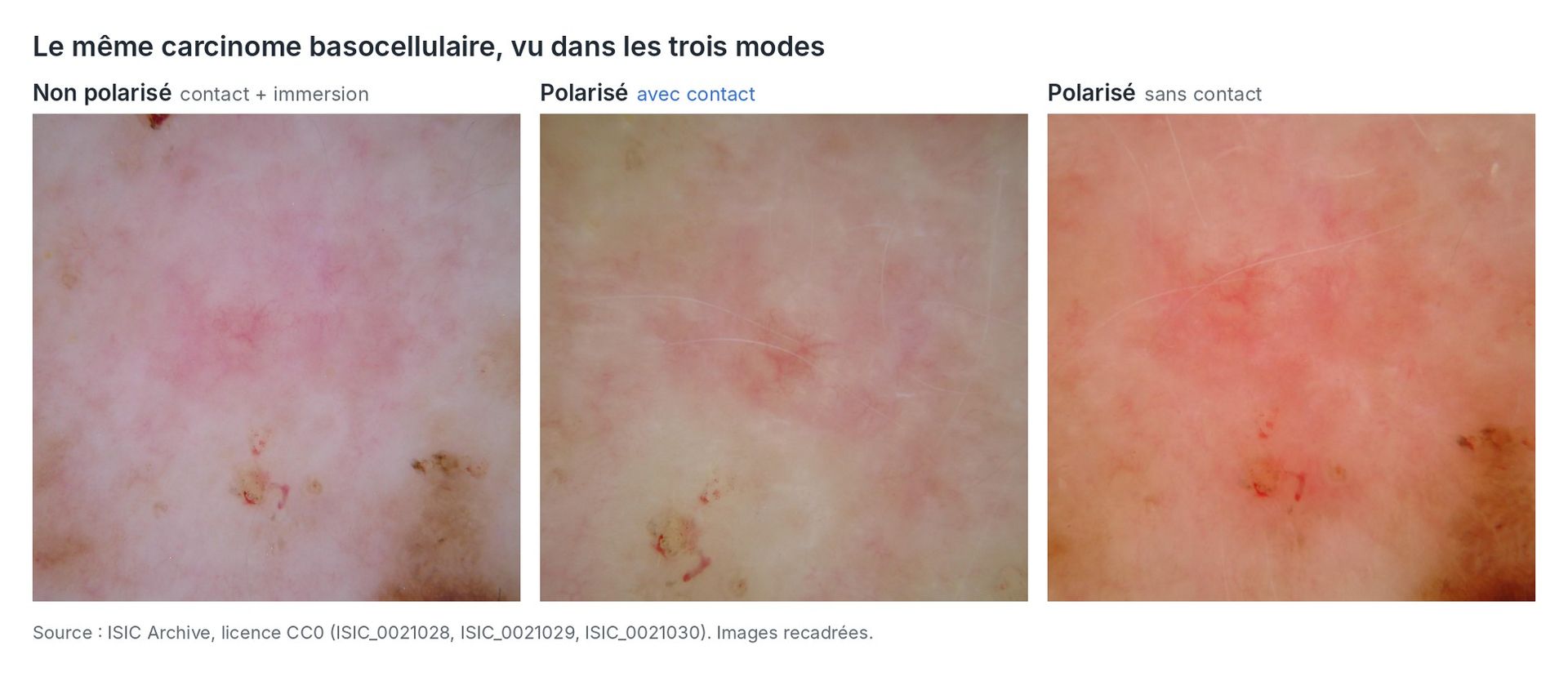
In pictures: the same basal cell carcinoma in all three modes. The three shots show the same lesion, with the same landmarks (small crust in the center, pigmentation at the bottom right). Yet the rendering changes markedly: without contact, no pressure is applied to the skin and pink dominates; in contact, the image is clearer and more uniform.
What the scientific literature says
Studies conclude that the modes agree strongly, with targeted differences that can affect diagnosis.
1. The overall patterns are the same. The Memorial Sloan-Kettering team compared the three modes on 90 lesions (55 melanocytic, 35 non-melanocytic). Agreement on global dermoscopic patterns was excellent (kappa 0.88 to 1.00). The differences mainly concerned the blue-white veil, pink color and milia-like cysts (Benvenuto-Andrade et al., 2007).
2. The difference is explained by depth. The same authors then demonstrated the physical origin of these differences: non-polarized light “reads” the superficial layers, polarized light the deeper layers. Collagen alters the polarization of light, which explains shiny white structures (Pan et al., 2008).
3. The chosen mode can change the diagnosis of beginners. In a pilot study, around a hundred dermatologists with little dermoscopy experience were asked about the same lesions seen in both modes. Diagnoses differed significantly for seborrheic keratosis, atypical nevus and melanoma (Wang et al., 2008).
4. Sensitivity versus specificity. Polarized light can increase sensitivity for amelanotic or structureless melanomas and basal cell carcinomas, by revealing vessels and white lines. Non-polarized light can increase specificity by identifying the milia-like cysts of seborrheic keratoses (dermoscopedia). For basal cell carcinomas, a dedicated study also describes differences in appearance between the two types of light (Liebman et al., 2012).
5. Not all “polarized” devices are equal. An international team photographed the same melanoma in situ with six dermatoscopes on the market. Some models clearly displayed the shiny white lines in polarized mode; others gave an image almost identical to non-polarized mode (Whybrew et al., 2022). The model in question was the Heine Delta 30. The manufacturer has corrected this on the Delta 30 Pro and UV, developed in collaboration with Dr Cliff Rosendahl, co-author of the study.
6. What about polarized with immersion? This is the least studied configuration in its own right: we found no clinical study comparing it directly with the other modes. Reference sources in the field nevertheless describe it as beneficial: the fluid is said to improve image quality in polarized contact mode (dermoscopedia, Polarized dermoscopy chapter). It is also essential for using both modes of a hybrid device (dermoscopedia).
How to choose for your practice
For most practitioners, a hybrid device (polarized + non-polarized) is the safest choice. The two modes are complementary, and combining them improves both diagnostic accuracy and practitioner confidence (DermNet).
A few additional pointers:
- in general practice, polarized contact mode offers a stable, sharp image, without the constraint of mandatory fluid. A drop of fluid improves the result further;
- non-contact remains a complement: convenient for a vascular lesion or a hard-to-reach area, but less reliable as a main mode;
- in podiatry, non-polarized light is reported to perform better on acral lesions, those on the soles of the feet (DermNet);
In summary
- Non-polarized with immersion: the skin surface, with precision. Ideal for milia-like cysts and the blue-white veil.
- Polarized contact: depth, with a stable image. Ideal for vessels and shiny white structures. A drop of fluid improves the image further.
- Polarized non-contact: a convenient complement, not a main mode.
- Both types of light together, in contact: the most complete information. That is the whole point of hybrid dermatoscopes.
Unsure between several models? Our team can advise you based on your practice.
References
- Benvenuto-Andrade C, Dusza SW, Agero AL, et al. Differences between polarized light dermoscopy and immersion contact dermoscopy for the evaluation of skin lesions. Arch Dermatol. 2007;143(3):329-338. doi:10.1001/archderm.143.3.329. Abstract
- Pan Y, Gareau DS, Scope A, Rajadhyaksha M, Mullani NA, Marghoob AA. Polarized and nonpolarized dermoscopy: the explanation for the observed differences. Arch Dermatol. 2008;144(6):828-829. doi:10.1001/archderm.144.6.828. Notice
- Wang SQ, Dusza SW, Scope A, Braun RP, Kopf AW, Marghoob AA. Differences in dermoscopic images from nonpolarized dermoscope and polarized dermoscope influence the diagnostic accuracy and confidence level: a pilot study. Dermatol Surg. 2008;34(10):1389-1395. doi:10.1111/j.1524-4725.2008.34293.x. Abstract
- Liebman TN, Jaimes-Lopez N, Balagula Y, et al. Dermoscopic features of basal cell carcinomas: differences in appearance under non-polarized and polarized light. Dermatol Surg. 2012;38(3):392-399. doi:10.1111/j.1524-4725.2011.02205.x
- Whybrew C, Pietkiewicz P, Kohut I, Chia JC, Akay BN, Rosendahl C. Not all polarized-light dermatoscopes may display diagnostically critical polarizing-specific features. Dermatol Pract Concept. 2022;12(4):e2022250. doi:10.5826/dpc.1204a250. Article
- Reiter O, Dimitriou F, Scope A, Marghoob AA, Braun RP. Differences between polarized and non polarized dermoscopy. dermoscopedia (International Dermoscopy Society). Chapter
- Thompson H, Oakley A. A comparison of polarised and nonpolarised dermoscopy. DermNet, 2020. Article
See also: Polarized dermoscopy. dermoscopedia (International Dermoscopy Society). Chapter