
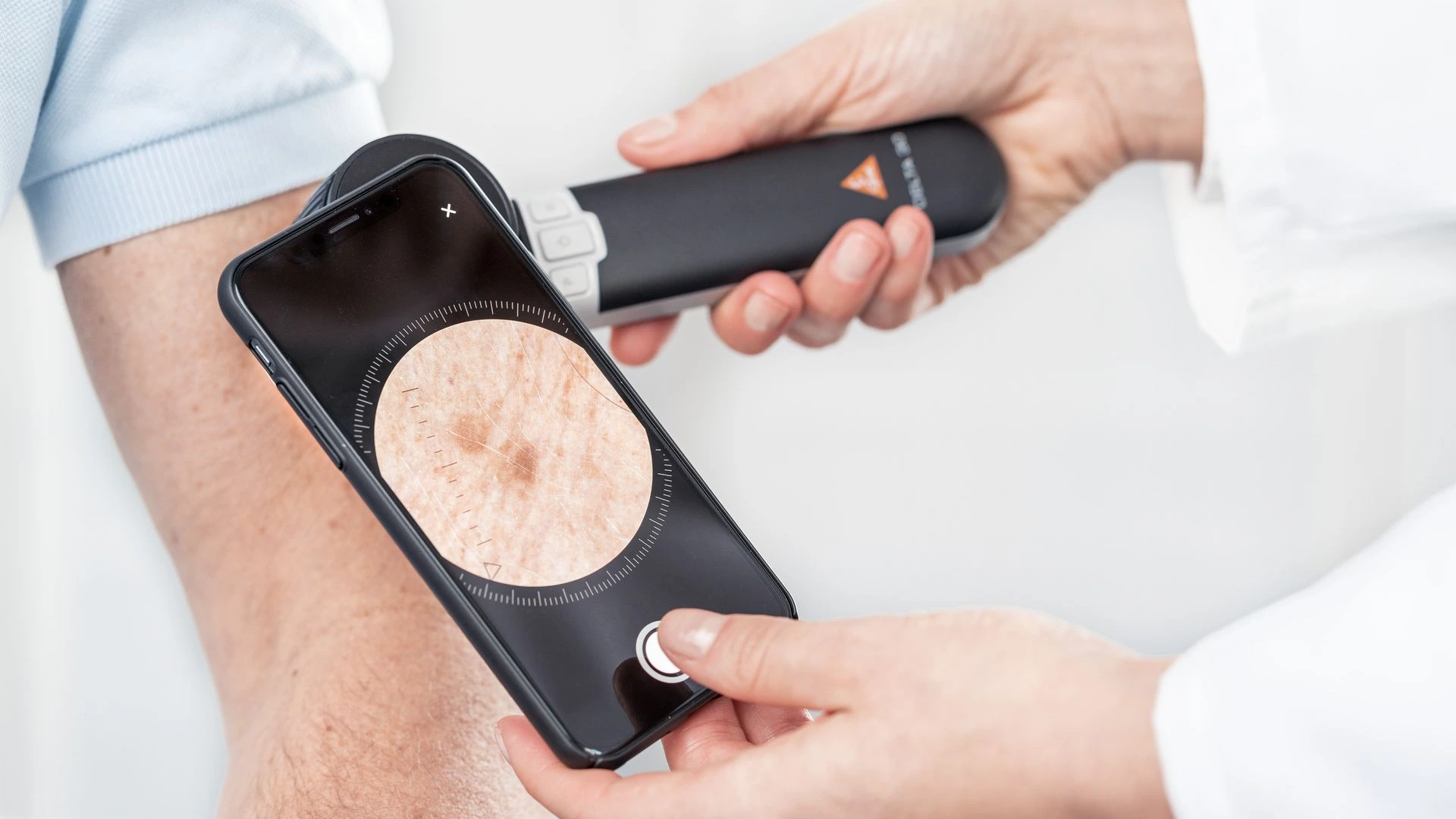
Attaching your dermatoscope to your smartphone lets you photograph in a few seconds what you see through the eyepiece. It is simple, inexpensive and very useful for following up a lesion or asking for an opinion. But a dermoscopic photo taken with a phone is not automatically a good medical image: color, sharpness and a rigorous shooting method make all the difference.
Key points in 30 seconds
- The principle: the dermatoscope creates the image (optics, lighting, polarization); the smartphone only records it.
- What the science says: dermoscopic images taken with a smartphone allow reliable triage of suspicious lesions and shorten the time to treatment of skin cancers.
- The limits: colors vary from one phone to another, there is no standardization, and patient photos end up in the personal photo gallery.
- Who it is for: ideal for documenting a lesion and asking for an opinion. For maximum quality or regular photographic follow-up, other solutions exist: we compare them in a dedicated article.
How it works
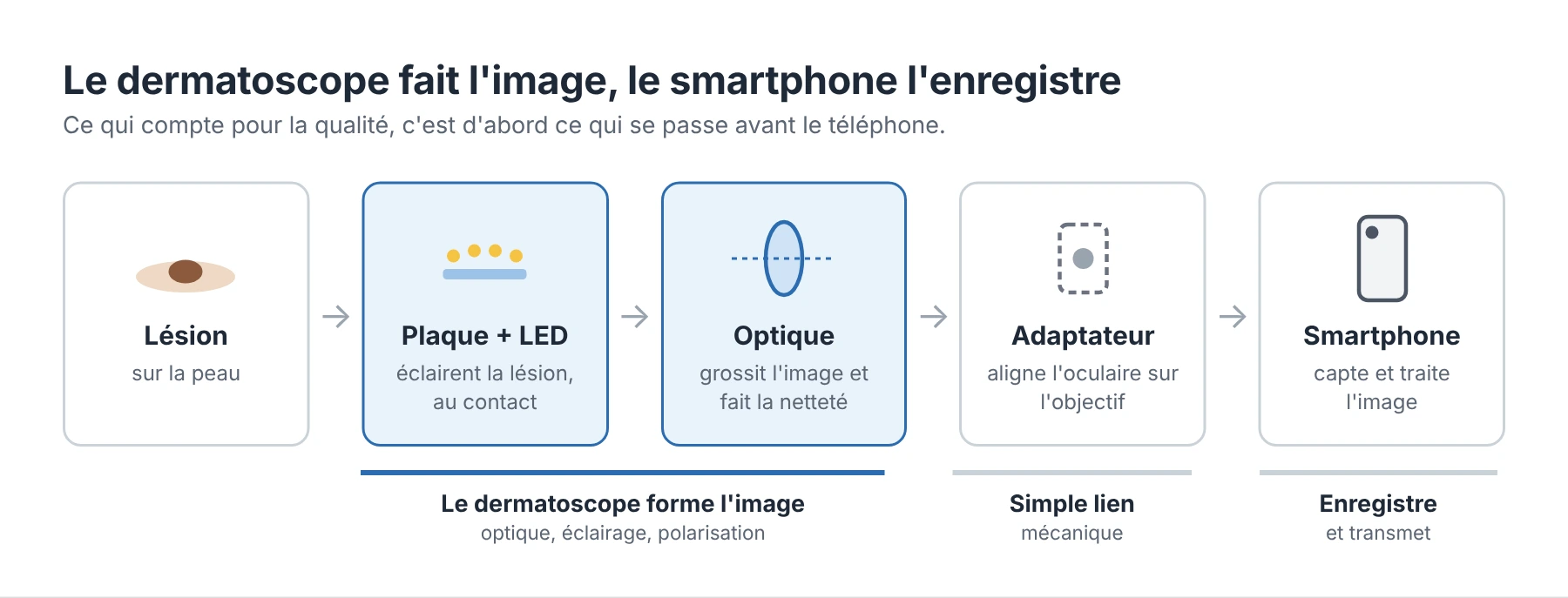
The dermatoscope creates the image; the smartphone only photographs it. The plate placed on the skin and the LEDs illuminate the lesion, the optics magnify it, and the adapter places the phone's lens in front of the eyepiece, where your eye would be.
In practice: a recent phone will not make up for a mediocre dermatoscope, whereas a good dermatoscope gives good images even with a mid-range phone.
The different types of adapters
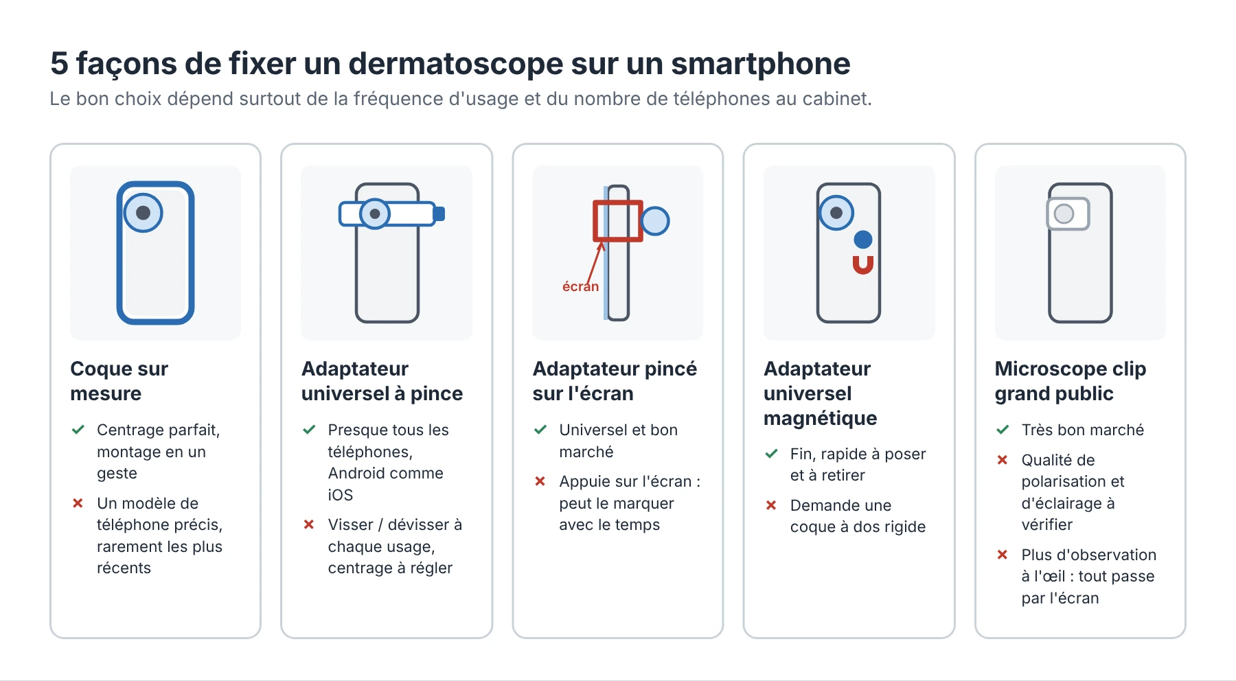
There are several ways to connect a dermatoscope to a smartphone. The right choice depends mainly on how often you use it and how many phones there are in the practice.
Type | Principle | Advantages | Points to watch |
|---|---|---|---|
Custom case | A case made for a specific phone model includes the dermatoscope mount | Perfectly centered on the lens, fitted in one move | Must be bought again with each new phone; often limited to iPhones, and rarely up to date for the latest models |
Universal clamp adapter | An adjustable holder clamps onto the phone, and the dermatoscope attaches to it magnetically | Compatible with almost all smartphones, Android and iOS | A little handling each time: screw to tighten, unscrew to remove. You also have to center the adapter yourself in front of the right lens |
Screen clip adapter | A clip sandwiches the phone, front and back, and holds the dermatoscope in front of the lens | Universal and inexpensive | The clip presses directly on the screen: over time, it can mark or damage it. Less stable mounting |
Universal magnetic adapter | A mounting button is stuck onto a hard case, and the dermatoscope clips onto it magnetically | Slim, quick to attach and remove, fits all camera layouts | Requires a hard-backed case (polycarbonate or acrylic) |
Consumer “clip-on” dermatoscope | A small magnifying lens attaches directly to the phone's lens | Generally inexpensive | Watch out for the quality of the polarization, and poorly controlled lighting and reflections. You also lose optical observation: the lesion can only be seen through the screen |
At HEINE, for example, the DELTA 30, DELTA 30 PRO and DELTAone connect to the smartphone with a universal magnetic adapter, which requires a hard-backed case. The brand also offers custom cases for iPhone models (the 12 Pro being the most recent) and a universal clamp adapter that holds the phone on the focusing ring.
The multi-lens phone trap: the adapter must be placed in front of the main lens. HEINE recommends testing each lens and keeping the one that gives a sharp image without vignetting. Do not use macro mode, as the image will be overexposed.
Image quality: what makes the difference
The quality of a dermoscopic photo depends first on the dermatoscope, then on the phone. A high-end smartphone will never make up for mediocre optics or poorly controlled lighting.
What comes from the dermatoscope
- The optics: magnification, field of view and no distortion at the edges.
- The lighting: uniform LEDs and stable light color, for accurate color rendering.
- The light mode: polarized, non-polarized and immersion do not show the same structures. For photography, we recommend polarized light in contact: the plate pressed on the skin stabilizes the distance and removes most of the glare.
- The graduated contact plate: the scale visible on the photo lets you measure the lesion and compare two pictures over time.
What comes from the smartphone
- Automatic image processing: phones “beautify” photos (contrast, saturation, smoothing). Nice for a portrait, a problem for a pigmented lesion.
- Automatic white balance and exposure, which change from one photo to the next.
- Digital zoom, which degrades the image instead of enlarging it.
- The choice of lens on phones with several cameras.
What the scientific studies say
The studies agree: a quality dermatoscope attached to a smartphone produces reliable images for triaging lesions and asking for an opinion. Dermoscopy adds real value compared with a simple phone photo.
Study | What was tested | Main result |
|---|---|---|
20 Swedish primary care centers referred their patients to a dermatologist via an app and a dermatoscope attached to a smartphone (816 patients), compared with 746 patients referred by paper letter | Much shorter time to surgery for melanomas and carcinomas; more than 40% of patients could have avoided a consultation; only 0.4% of images unusable | |
113 skin tumors photographed with a phone, with a dermatoscope placed on the lens, then assessed remotely | Very good agreement with the reference diagnosis (κ = 0.84); but 6% of dermoscopic images unusable, versus 1% of clinical photos | |
Systematic review of teledermatology for diagnosing skin cancer | Fewer than 7% of malignant lesions missed; considered a useful tool to help general practitioners decide whom to refer | |
Skin carcinomas: dermoscopic images vs simple smartphone photos, analyzed by artificial intelligence | Accuracy of 87.8% with dermoscopy versus 74.8% with the smartphone photo alone | |
Inexpensive “clip-on” microscope (Nurugo) compared with a conventional dermatoscope | Encouraging results on 78 lesions, but the authors themselves list the shortcomings: flash reflections, no polarized light, a field of view of only 3 mm², non-medical device |
Key takeaways
- Smartphone dermoscopy is a validated triage and communication tool, particularly between general practitioners and dermatologists.
- Image quality remains the weak point: the share of unusable photos fell sharply between 2011 and 2015, thanks to better devices and a shooting protocol.
- A smartphone photo without a dermatoscope performs much worse than a dermoscopic image.
- A consumer gadget does not replace a medical dermatoscope.
Limits to keep in mind
The smartphone was designed to take beautiful photos, not accurate ones. This is the main limit of mobile dermoscopy, and it has four practical consequences.
1. Colors that vary from one phone to another. In dermoscopy, color is a diagnostic criterion (blue-gray, brown, red…). Yet each phone interprets colors in its own way, and its automatic settings change from one photo to the next. A study of a pocket dermatoscope attached to a smartphone shows that color calibration brings the color difference down to a level close to what the eye perceives (Cugmas et al., 2017). Without calibration, nothing guarantees it.
2. Follow-up over time is hard to standardize. Comparing a lesion six months apart requires the same framing, the same light and the same rendering. With a phone that updates itself, or that is replaced every two or three years, these conditions are no longer met.
3. A single lesion, not a full examination. A transmitted photo shows only the chosen lesion. The authors of the studies point out that teledermoscopy does not replace a full skin examination, during which other lesions are often discovered.
4. Health data on a personal phone. A photo of a lesion is health data, for which the practitioner is responsible under the GDPR. It should neither stay in the personal photo gallery nor be shared through a consumer messaging app (CNIL). Transfer, archiving and sending for teledermatology will be the subject of a future article.
Good practice for a usable image
A few habits are enough to turn a “decent” photo into an image that is useful to a colleague.
- Always two photos: an overview to locate the lesion on the body, then the dermoscopic view.
- Work in contact, with polarized light, with the plate cleaned and disinfected between patients.
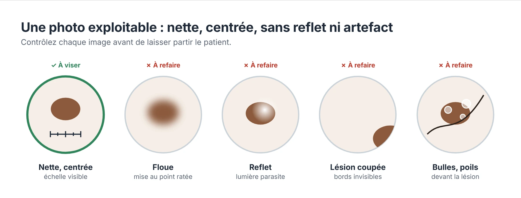
- Center the lesion and keep it whole in the field, with the graduated scale visible.
- Focus on the dermatoscope, then tap the screen to lock the phone's focus and exposure.
- Do not use digital zoom or filters, and turn off “beauty” or portrait modes.
- Always use the same phone and the same dermatoscope to follow the same lesion over time.
- Check the photo before the patient leaves: sharp, with no bubbles, glare or hair in front of the lesion.
- Store and transmit securely, via a medical photography or teledermatology app, never in the personal photo gallery.
Further reading
The smartphone is the gateway to dermoscopic photography. If you take photos often, follow patients over time or are looking for the best possible quality, a DSLR or mirrorless camera, or a dedicated camera such as the Casio DZ-D100, may be more suitable.
Photography in dermoscopy: smartphone, camera or dedicated device?
References
Scientific studies
- Börve A, Dahlén Gyllencreutz J, Terstappen K, et al. Smartphone teledermoscopy referrals: a novel process for improved triage of skin cancer patients. Acta Derm Venereol. 2015;95(2). doi:10.2340/00015555-1906
- Börve A, Terstappen K, Sandberg C, Paoli J. Mobile teledermoscopy — there's an app for that! Dermatol Pract Concept. 2013.
- Kroemer S, Frühauf J, Campbell TM, et al. Mobile teledermatology for skin tumour screening: diagnostic accuracy of clinical and dermoscopic image tele-evaluation using cellular phones. Br J Dermatol. 2011;164(5):973-979.
- Chuchu N, Dinnes J, Takwoingi Y, et al. Teledermatology for diagnosing skin cancer in adults. Cochrane Database Syst Rev. 2018;12:CD013193.
- Dascalu A, Walker BN, Oron Y, David EO. Non-melanoma skin cancer diagnosis: a comparison between dermoscopic and smartphone images by unified visual and sonification deep learning algorithms. J Cancer Res Clin Oncol. 2021. doi:10.1007/s00432-021-03809-x
- Veronese F, Branciforti F, Zavattaro E, et al. The role in teledermoscopy of an inexpensive and easy-to-use smartphone device for the classification of three types of skin lesions using convolutional neural networks. Diagnostics. 2021;11(3):451.
- Cugmas B, Pernuš F, Likar B. Color constancy in dermatoscopy with smartphone. Proc SPIE 10592, Biophotonics—Riga 2017.
Recommendations and documentation