
This article is an informational summary of the scientific literature. It does not replace training or the practitioner's clinical judgment
UV light, a new tool for the dermatoscope
In three years, UV dermoscopy has gone from a curiosity to a tool studied in journals such as JAAD, JEADV and Dermatology and Therapy. Publications from 2025 and 2026 confirm that it does not replace polarized dermoscopy, but that it provides additional clues.
The principle is not new: the Wood's lamp has illuminated the skin with UV light for more than a century. What is new is building this light into a pocket dermatoscope, with magnification. The lesion can thus be examined in polarized light and then in UV, in a single step.
This article reviews what the studies show, then gives the full list of references, sorted by topic.
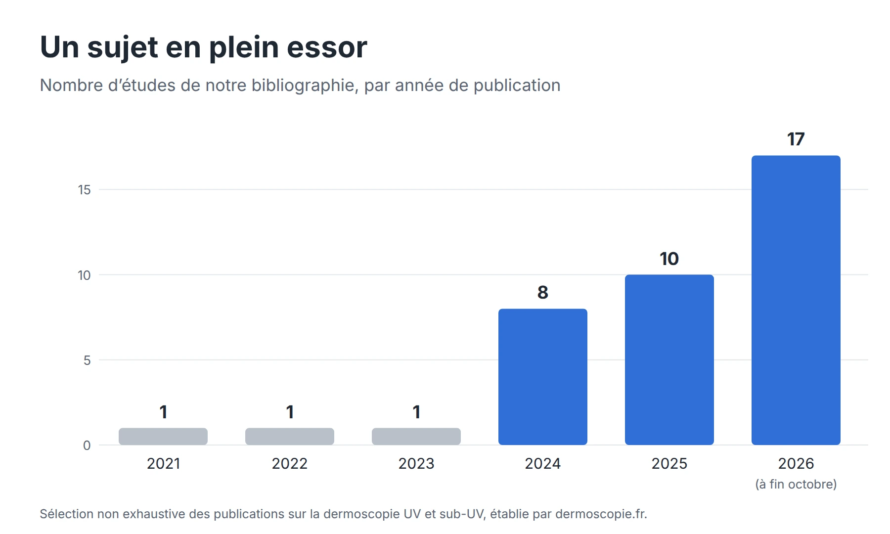
Number of studies on UV dermoscopy per year, 2021 to 2026
Two ways to use UV
The literature distinguishes two modalities, which do not show the same thing. The systematic review by Pietkiewicz et al. (2024) proposed a common terminology for both.
Modality | Wavelength | What you see | Main uses studied |
|---|---|---|---|
UV-induced fluorescence dermoscopy (UVFD) | 365 nm | Fluorescence emitted by the skin (keratin, porphyrins, fungi, parasites) | Keratinizing tumors, basal cell carcinoma, melanoma, scabies, fungal infections, Demodex |
Sub-UV reflectance dermoscopy (sUVRD), or violet light | About 405 nm | Reflected light, strongly absorbed by melanin: the pigment stands out clearly | Melanocytic lesions, pigment borders, melanonychia, acral melanoma |
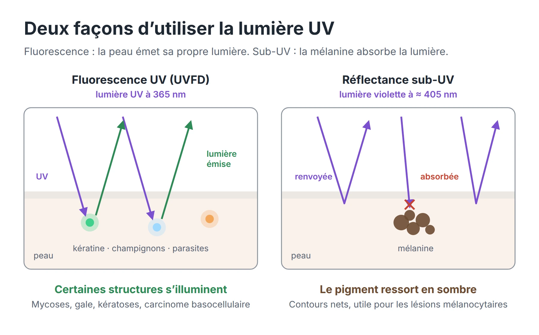
Diagram: UV fluorescence at 365 nm and sub-UV reflectance at 405 nm on the skin
In both cases, UV examination complements polarized dermoscopy. Polarized light remains the reference for vessels and most pigmented structures.
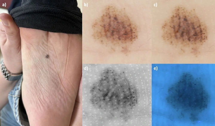
Acral nevus of the sole: (a) clinical photo, (b) polarized dermoscopy, (c) non-polarized, (d) sub-UV reflectance, which highlights the openings of the eccrine ducts, (e) UV fluorescence.
Source: Erol Mart HM, Aydemir AT, Akay BN, Pietkiewicz P. Int J Dermatol. 2026;65:1625-1632. doi:10.1111/ijd.70384. CC BY 4.0 license.
Skin tumors and melanoma: what the studies show
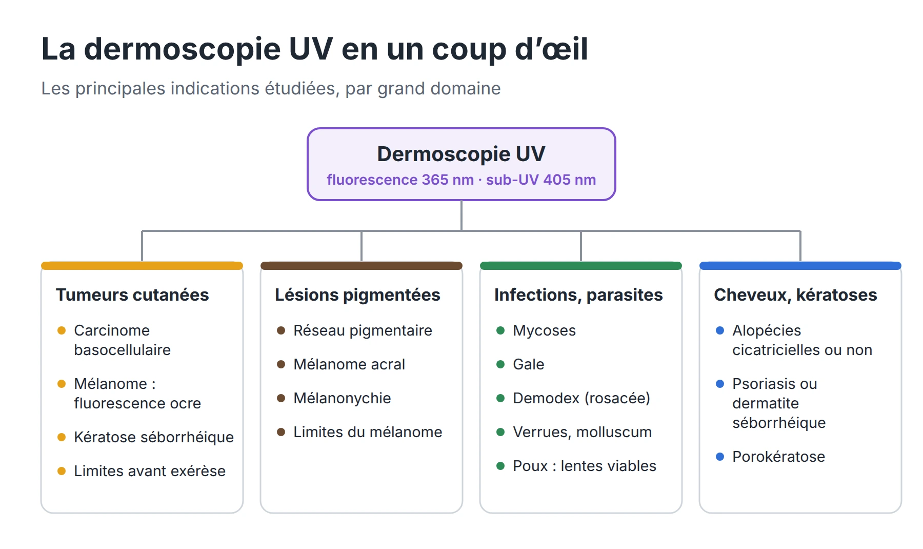
Keratin-related structures stand out better under UV. A multicenter study published in JAAD (Navarrete-Dechent et al., 551 lesions) concludes that UVFD better highlights keratin, comedo-like openings and the cerebriform pattern (“fissures and ridges”). Lesion borders are also better delineated.
“Ochre fluorescence” is a clue to invasive melanoma. An international study (JAAD, 2026, 234 lesions) found this ochre fluorescence in 23% of melanomas versus 3% of other lesions. It was much more frequent in invasive melanomas than in melanomas in situ (40% vs 6%). Its specificity is 97%, but its sensitivity is only 23%: its presence is a warning sign, its absence is not reassuring. Several 2026 publications extend this line of research: a case series, an article on its absence in acral melanoma and a hypothesis on its physicochemical origin.
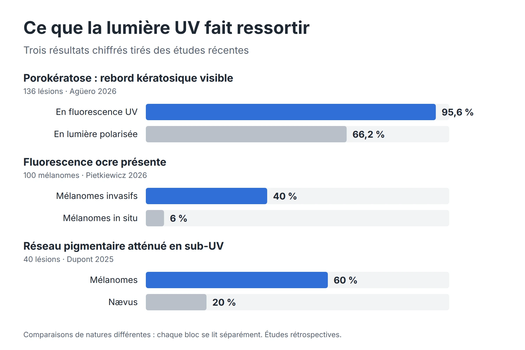
Chart: porokeratosis, ochre fluorescence and pigment network, figures compared
For basal cell carcinoma, facial lesions and excision margins:
- In 163 basal cell carcinomas, UVFD provides the most clues for small tumors under 5 mm, facial lesions and nodular or non-pigmented forms (Wojtowicz and Żychowska, 2024). A second study of 151 head and neck carcinomas confirms these signs, in particular dark silhouettes (Wojtowicz et al., 2025).
- In 181 non-pigmented facial lesions, dark silhouettes appear in 84% of basal cell carcinomas. Several lesion-specific signs help distinguish them from sebaceous hyperplasia, dermal nevus and seborrheic keratosis (Pszczołkowska et al., 2026).
- The multiple aggregated yellow-white globules of basal cell carcinoma, which are easy to miss, become fluorescent and therefore more visible with UVFD.
- UV light also helps delineate tumor margins before excision (the “umbrella effect” described under Wood's light).
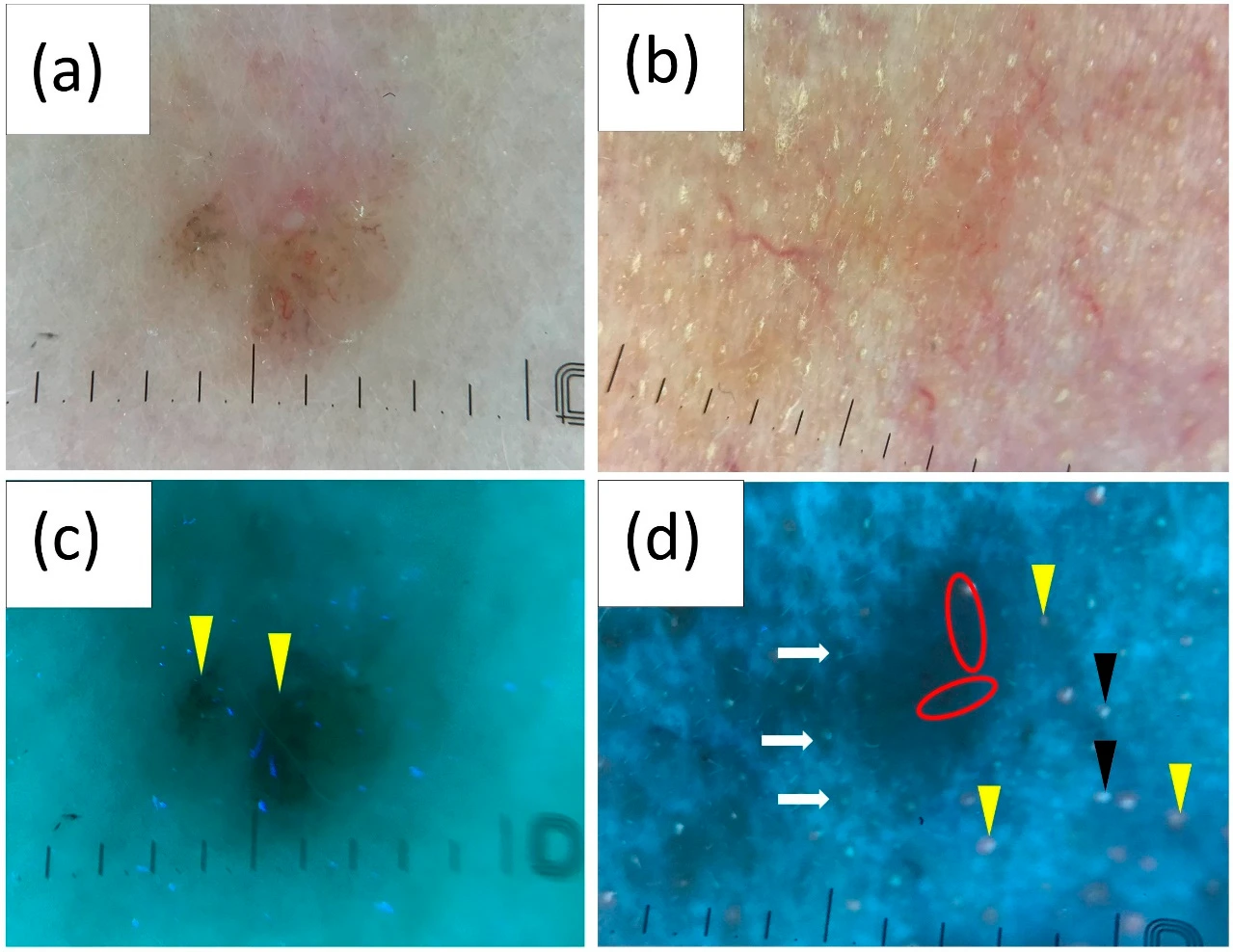
Pigmented (a) and non-pigmented (b) basal cell carcinoma in polarized dermoscopy, and their UV fluorescence images (c, d). The black globules (c) and the interrupted follicular pattern (d) stand out under UV.
Source: Wojtowicz I, Żychowska M. Application of Ultraviolet-Enhanced Fluorescence Dermoscopy in Basal Cell Carcinoma. Cancers. 2024;16:2685. doi:10.3390/cancers16152685. CC BY 4.0 license.
Sub-UV light is useful for melanocytic lesions:
- At the Centre Léon Bérard (Lyon), the visibility of the pigment network decreased under sub-UV light in 60% of melanomas, versus 20% of nevi (Dupont et al., 2025).
- Several Japanese teams have shown that 405 nm light sharpens the distribution of melanin, which helps delineate melanoma.
- In 42 acral lesions, conventional dermoscopy remains superior for most structures, but sub-UV light better shows the openings of the eccrine ducts (Erol Mart et al., 2026). Longitudinal melanonychia was also studied under sub-UV light in 2026.
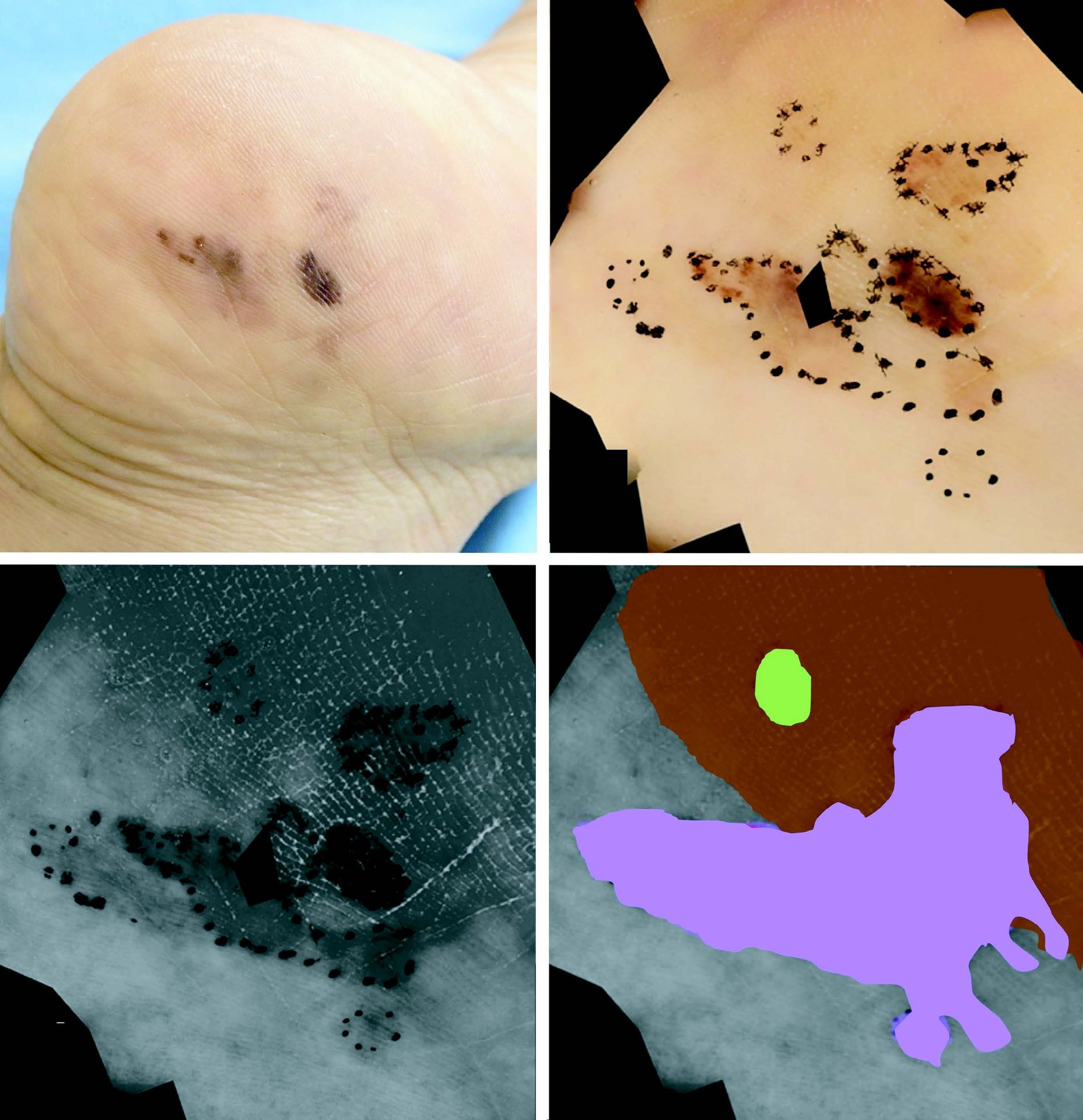
Heel melanoma: clinical photo (a), conventional dermoscopy (b) and violet light (c). Violet light reveals an extension of the pigment that conventional dermoscopy did not show.
Source: Shu J, Yamamoto Y, Aoyama K, Togawa Y, Kishimoto T, Matsue H. J Dermatol. 2022;49:710-713. doi:10.1111/1346-8138.16389. CC BY 4.0 license.
General dermatology: infections, infestations, keratoses
This is the field where UV dermoscopy has the clearest results. It takes over the historical indications of the Wood's lamp, with magnification added.
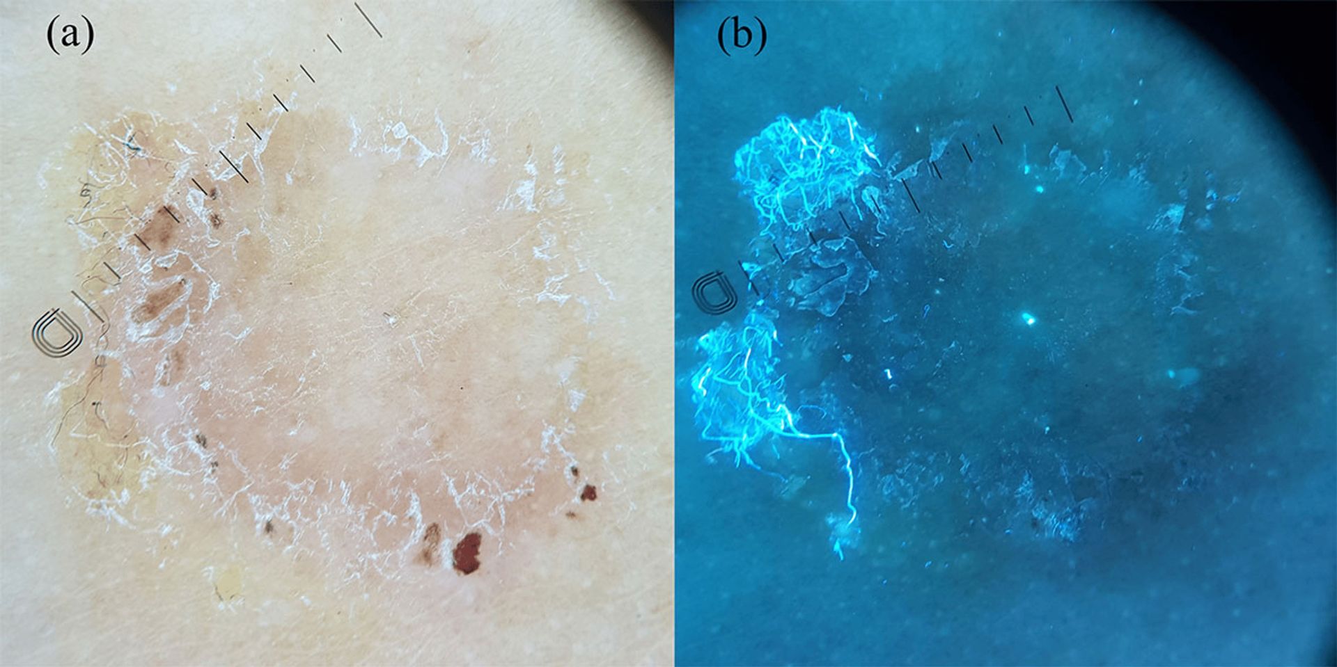
Tinea corporis: in polarized dermoscopy (a), an annular lesion; in UV fluorescence (b), blue-green fluorescence on the active border.
Source: Elbadawy et al. Diagnostic Value of Ultraviolet-Induced Fluorescence Dermoscopy in Cutaneous Fungal Infection. Cureus. 2026;18(6):e110437. doi:10.7759/cureus.110437. CC BY 4.0 license.
Indication | Key result | Study |
|---|---|---|
Cutaneous fungal infections | Sensitivity of 98.6% and specificity of 80.5% compared with direct KOH examination, in 150 patients | Cureus, 2026 |
Scabies | Better detection of burrows and eggs on dark skin, in 97 lesions and 43 patients. The “ball sign” shows the body of the mite | Errichetti et al., 2024; Kaliyadan et al. |
Demodex (rosacea) | Above 11 bright blue dots, sensitivity of 82% and specificity of 71% for the presence of Demodex, compared with skin surface biopsy | Clin Exp Dermatol, 2026 |
Porokeratosis | Peripheral keratotic rim visible in 95.6% of cases with UVFD, versus 66.2% in polarized light, in 136 lesions | Agüero et al., 2026 |
Seborrheic keratosis | Comedo-like openings and cerebriform pattern better highlighted under near-UV light, in 207 pigmented lesions | Acta Derm Venereol, 2023 |
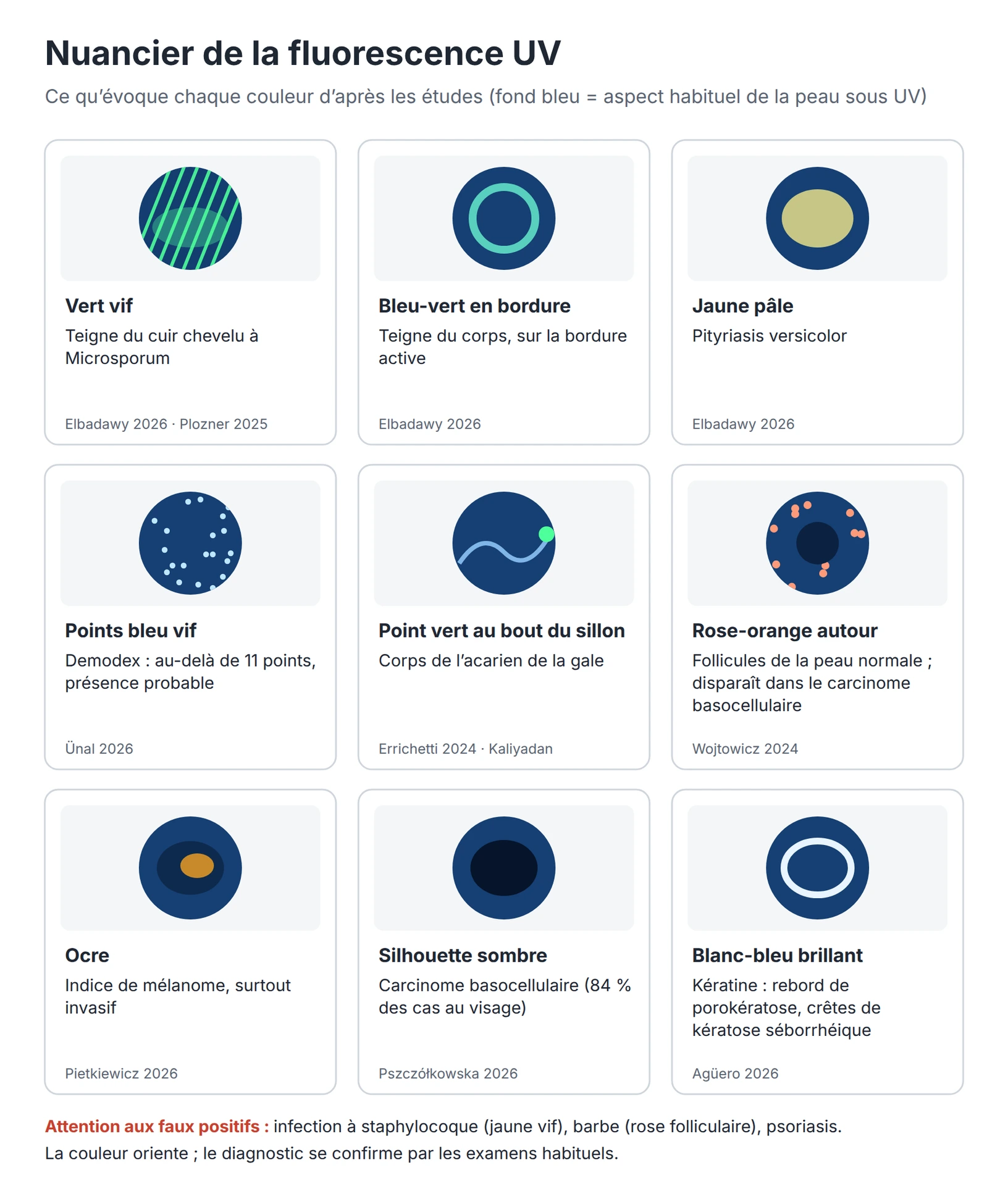
Color chart of UV fluorescence colors and what they suggest
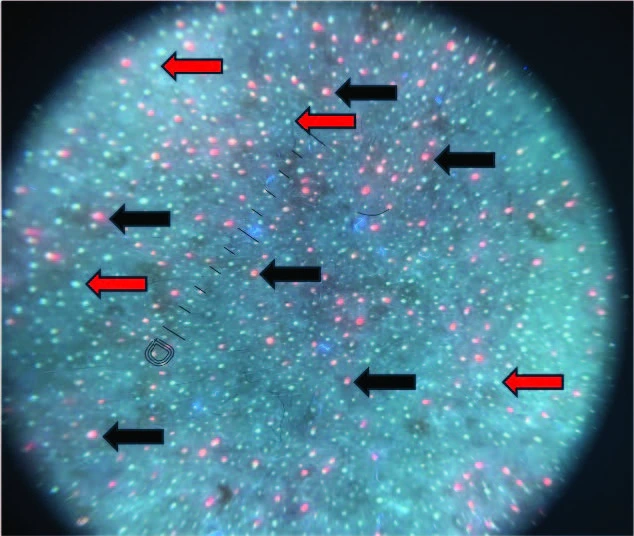
Rosacea in UV dermoscopy: numerous fluorescent dots, of two types (arrows). Above 11 bright blue dots, the presence of Demodex is likely.
Source: Ünal E, Yücel MB, Yiğit M, Yaman O, Ertaş R, Akay BN. Clin Exp Dermatol. 2026;51:1400-1405. doi:10.1093/ced/llag100. CC BY 4.0 license.
For the scalp, UVFD distinguishes scarring from non-scarring alopecias better than polarized light, with better interobserver agreement (kappa of 0.83 vs 0.57, in 84 patients). It also helps differentiate psoriasis from seborrheic dermatitis. In Microsporum tinea it reveals green fluorescence, and in pediculosis it shows viable nits. Other 2026 studies cover warts, molluscum contagiosum and mite viability in scabies.
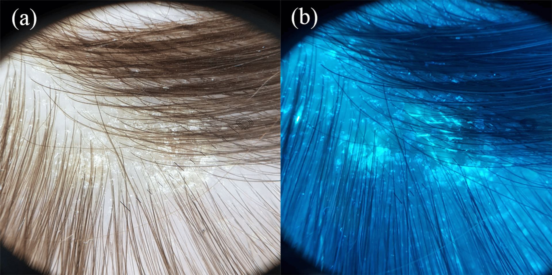
Tinea capitis: perifollicular scales in polarized dermoscopy (a); bright green fluorescence under UV (b).
Source: Elbadawy et al., Cureus, 2026. doi:10.7759/cureus.110437. CC BY 4.0 license.
Limits to keep in mind
All the authors present UV dermoscopy as a complement, never as a stand-alone examination.
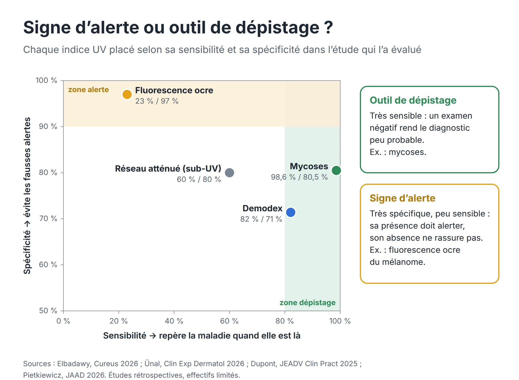
Chart of sensitivity and specificity of four UV dermoscopy clues
- Most studies are retrospective, involve few patients and are conducted in a single center.
- Some signs are highly specific but not very sensitive. For example, ochre fluorescence is absent in 77% of melanomas.
- Polarized light remains superior for vessels, and sub-UV light visualizes deep lesions poorly.
- Terminology is not yet fully harmonized, and the rendering may vary with the device and the camera sensor.
Bibliography: studies on UV dermoscopy
This list brings together publications from 2020 to 2026, sorted by topic and from most recent to oldest.
Reviews and summaries
- Trecarichi et al. Dermoscopy in the Treatment Monitoring of Cutaneous Infectious Diseases: A Review. Dermatologic Therapy, 2026. Link
- Ultraviolet-Induced Fluorescence Dermoscopy: Game Changers. J Clin Aesthet Dermatol, 2025. Link
- Plozner N, Bhat YJ, Errichetti E. Applications of UV-Induced Fluorescence (UVF) Dermoscopy in Infectious Dermatoses (UVF-Infectiouscopy). Dermatol Pract Concept, 2025. Link
- Bhat et al. Ultraviolet-Induced Fluorescence Dermoscopy, a Novel Diagnostic Technique in Dermatological Practice: A Systematic Review. Indian Dermatol Online J, 2025. Link
- Ioannides D, Papadimitriou I. Ultraviolet-induced fluorescence dermoscopy improves the differential diagnosis in the field of general dermatology. JEADV, 2025 (commentary). Link
- Pietkiewicz P et al. Applications of Ultraviolet and Sub-ultraviolet Dermatoscopy in Neoplastic and Non-neoplastic Dermatoses: A Systematic Review. Dermatol Ther (Heidelb), 2024. Link
- Kaliyadan F et al. Ultraviolet reflectance dermoscopy. Indian J Dermatol Venereol Leprol, 2024. Link
Skin tumors and melanoma (UV fluorescence)
- Pietkiewicz P et al. Ochre fluorescence under ultraviolet-induced fluorescence dermatoscopy is a strong clue to melanoma. JAAD, 2026. Link
- Meduri et al. Ochre Fluorescence in Melanoma Under Ultraviolet-Induced Fluorescence Dermoscopy: A Case Series. Int J Dermatol, 2026. Link
- Mart et al. Absence of Ochre Fluorescence in Acral Melanoma Under Ultraviolet Dermoscopy. Int J Dermatol, 2026. Link
- On the likely physicochemical basis of the “ochre color” under ultraviolet-induced fluorescent dermoscopy. 2026. Link
- Pszczołkowska, Reich, Żychowska. Ultraviolet-Induced Fluorescence Dermoscopy for the Differentiation of Non-Pigmented Malignant and Benign Lesions on the Face. Dermatol Ther (Heidelb), 2026. Link
- Navarrete-Dechent C et al. Ultraviolet-induced fluorescent dermoscopy for the diagnosis of skin tumors: A multicenter study. JAAD, 2025. Link
- Wojtowicz et al. Polarized Dermoscopy and Ultraviolet-Induced Fluorescence Dermoscopy of Basal Cell Carcinomas in the H- and Non-H-Zones of the Head and Neck. Dermatol Ther (Heidelb), 2025. Link
- Wojtowicz I, Żychowska M. Application of Ultraviolet-Enhanced Fluorescence Dermoscopy in Basal Cell Carcinoma. Cancers, 2024. Link
- Navarrete-Dechent C et al. The role of ultraviolet-induced fluorescence dermatoscopy for the detection of multiple aggregated yellow-white globules in basal cell carcinoma. JAAD, 2024 (letter).
- Ultraviolet induced fluorescence dermoscopy in seborrheic keratosis structures. JAAD, December 2024 (research letter).
- Gil-Pallares P et al. Identification of skin tumors margins using Wood’s light: “the umbrella effect”. Photodiagnosis Photodyn Ther, 2024. Link
Melanocytic lesions (sub-UV and violet light)
- Mart et al. Sub-ultraviolet reflectance dermoscopy in longitudinal melanonychia. JEADV, 2026. Link
- Erol Mart HM, Akay BN, Pietkiewicz P et al. Acral Melanocytic Lesions Under Conventional, Sub-Ultraviolet Reflectance, and Ultraviolet-Induced Fluorescence Dermoscopy: A Comparative Analysis. Int J Dermatol, 2026. Link
- Anriot J, Amini-Adle M (Centre Léon Bérard) et al. Sub-Ultraviolet Reflectance and Ultraviolet-induced Fluorescence Dermatoscopy in Action: Shedding Light on a Case of Balloon Cell Melanoma. Dermatol Pract Concept, 2026. Link
- Sub-UV reflectance dermatoscopy to reveal subtle dermatoscopic features: A case report. 2026. Link
- Dupont et al. Reduced Visibility of Pigment Network Under Sub-UV Reflectance Dermatoscopy. JEADV Clinical Practice, 2025. Link
- Caposiena Caro RD, Zalaudek I et al. Dermoscopy with subultraviolet light to distinguish the depth of melanocytes. JAAD, 2025. Link
- Near-ultraviolet Light Dermoscopy for Identification of Pigmented Skin Tumours. Acta Derm Venereol, 2023. Link
- Shu J, Togawa Y et al. Assessment of malignant melanoma lesions using violet-light dermoscopy: A case report. J Dermatol, 2022. Link
- Sano T, Minagawa A et al. Dermoscopy with near-ultraviolet light highlights the demarcation of melanin distribution in cutaneous melanoma. JAAD, 2021. Link
Infections, infestations and general dermatology
- Diagnostic Value of Ultraviolet-Induced Fluorescence Dermoscopy in Cutaneous Fungal Infection. Cureus, 2026. Link
- Ünal E, Akay BN et al. Detection of Demodex in rosacea: standardized skin surface biopsy vs. ultraviolet dermoscopy. Clin Exp Dermatol, 2026. Link
- Agüero, Navarrete-Dechent et al. Ultraviolet-Induced Fluorescence Dermatoscopy (UVFD) features in Porokeratosis: Findings from a Multicenter Retrospective Study. Dermatol Pract Concept, 2026. Link
- Characterizing UV-induced fluorescence dermatoscopy (UVFD) of warts and molluscum contagiosum and its utility for expert and novice dermatoscopists. Arch Dermatol Res, 2026. Link
- Magnified ultraviolet-induced fluorescence dermatoscopy for assessing mite viability in scabies. 2026. Link
- Combining fluorescent dye with ultraviolet dermoscopy can enhance visualization of porokeratosis and burrows of scabies. 2026. Link
- Kaliyadan F et al. The ‘ball sign’ in UV dermoscopy of scabies. Indian J Dermatol Venereol Leprol. Link
- Plozner N, Enechukwu NA, Bhat YJ, Behera B, Pietkiewicz P, Errichetti E. Diagnostic Accuracy of Polarized and Ultraviolet Fluorescence-Induced Dermoscopy in Scarring and Nonscarring Alopecias: a Retrospective Observational Multicentric Study. Dermatol Ther (Heidelb), 2025. Link
- Kołcz K, Reich A, Żychowska M. Application of Ultraviolet-Induced Fluorescence Trichoscopy (UVFT) in Hair and Scalp Diseases. Dermatol Ther (Heidelb), 2025. Link
- Errichetti E, Plozner N et al. Dermoscopy of Scabies: Utility of Polarised and Ultraviolet-Induced Fluorescence Examination in Fair and Dark Skin. Australas J Dermatol, 2024. Link
- Errichetti E, Pietkiewicz P, Bhat Y et al. Diagnostic accuracy of ultraviolet-induced fluorescence dermoscopy in non-neoplastic dermatoses (general dermatology): A multicentric retrospective comparative study. JEADV, 2024. Link
Technology and artificial intelligence
- Deep learning based hair removal on ultraviolet-induced fluorescence dermatoscopy images. Comput Methods Programs Biomed, 2026. Link
We do our best to keep it up to date as new publications on the subject appear.
Do you know of a study that is not listed?
Please let us know.
In practice
UV dermoscopy adds to the usual routine: you first examine the lesion in polarized light, then switch to UV mode to look for an additional clue. The strongest data today concern fungal infections, scabies, keratinizing lesions and decision support for an equivocal melanocytic lesion.
Would you like to move to a dermatoscope with a UV mode or sub-UV light? Discover our range:
UV mode (365 nm):
Sub-UV mode (405 nm):