
The dermatoscope has become the basic tool of skin examination, in dermatology practices as well as in general practice and podiatry. But between a pocket model at under €500 and a dermatology camera at over €2,500, the differences are not obvious. This guide covers the criteria that really change the examination, then compares the models one by one, without naming a “best” one: the right choice depends on your practice.
Key points in 30 seconds
- Dermoscopy clearly improves melanoma diagnosis, provided you are trained.
- Criterion no. 1: being able to switch from polarized to non-polarized light, and to examine in polarized contact mode.
- Next come the field of view (14 to 30 mm), photography, UV and battery life.
- The best way to choose: try it. A demo loan is available.
Why get equipped: what the studies say
Added to naked-eye examination, dermoscopy clearly improves melanoma diagnosis. In the 2018 Cochrane review (26 studies, more than 23,000 lesions examined in clinical practice), sensitivity rises from 76% to 92% at equal specificity (80%), and specificity from 75% to 95% at equal sensitivity (80%) [1]. An older meta-analysis, limited to studies in real-life conditions, reaches the same conclusion [2].
In general practice, a randomized trial of 73 physicians in Spain and Italy showed that after one day of training, dermoscopy raised the sensitivity of triaging suspicious lesions from 54.1% to 79.2%, with no loss of specificity (71.3% vs 71.8%) [3].
Two limits to keep in mind: the benefit is greater the more experienced the user [1], and the device does not replace training. Choosing a dermatoscope therefore comes second, after deciding to get trained.
The six criteria that matter
1. Polarized light, non-polarized light or immersion
This is the most important criterion, because the two types of lighting do not show the same things [4, 5]:
- Non-polarized, in contact with an immersion fluid: it better shows the skin surface (milia-like cysts, comedo-like openings of seborrheic keratoses, blue-white veil).
- Polarized: it removes glare without fluid (a drop of fluid improves the image further) and better shows deeper structures, vessels and above all shiny white structures (formerly “crystalline structures”), which are visible only in polarized light.
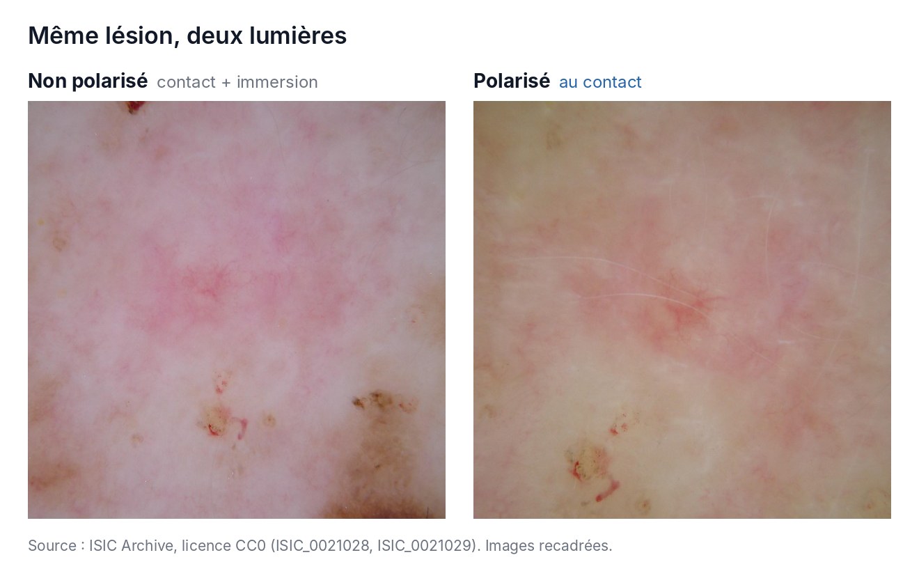
These shiny white structures matter: in a meta-analysis of 40 studies (22,796 lesions), they show a specificity of 93.6% for melanoma, with a sensitivity of 30.5% [6]. In other words, they are rare, but highly suggestive when present.
In practice, polarized contact mode (contact plate placed on the skin) is the one we recommend for routine examination: the image is stable, sharp and glare-free. Polarized dermoscopy with immersion fluid has not been the subject of a dedicated clinical study, but dermoscopedia describes it favorably [5].
All the models in this comparison, except the mini 3000, switch from polarized to non-polarized mode at the press of a button, which lets you compare both images of the same lesion. Note: not all polarized dermatoscopes render these structures in the same way. A 2022 research letter photographed the same melanoma in situ with six dermatoscopes: five showed the shiny white structures, the HEINE DELTA 30 did not [7]. This is a single case, but it is a good reason to try the device before buying it. According to the manufacturer, this has been corrected on the DELTA 30 PRO and DELTA 30 UV, whose LEDs have been repositioned [8], in collaboration with Dr Cliff Rosendahl.
Further reading: our article Polarized, non-polarized or immersion light: what is the difference?.
2. Field of view and magnification
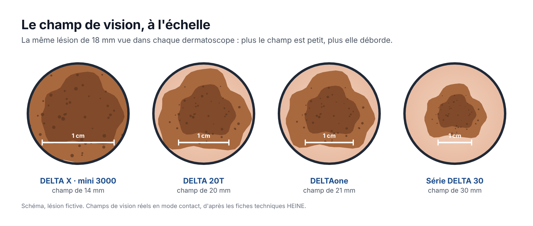
Standard magnification is 10x. What varies most is the field of view: from 14 mm on the DELTA X and the mini 3000 to 30 mm on the DELTA 30 series. A large field lets you see a large lesion in full and speeds up total body examination; a small head is easier to place behind the ear or between the toes. Most HEINE models accept a small contact plate for these hard-to-reach areas.
3. Contact or non-contact
Contact examination stabilizes the image and the graduated contact plate lets you measure the lesion: this is the mode to favor day to day. Without contact, vessels are not compressed and hygiene is simpler, but the image is more sensitive to movement and stray reflections: it is a complement rather than a main mode. On HEINE models, you simply remove the contact plate to switch from one to the other; the DELTA X also offers single-use or reusable polymer contact plates. The HEINE mini 3000 is used in contact only, with an immersion fluid; the Casio DZ-S50 only without contact.
4. Photo documentation
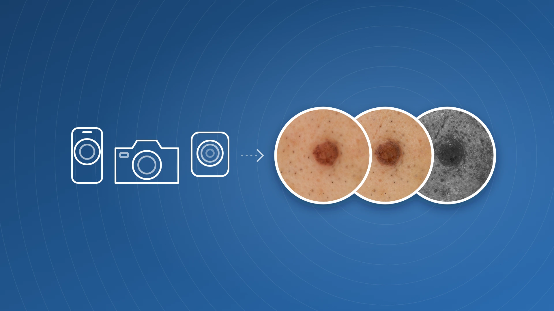
For patient follow-up or teledermatology, you need to be able to photograph what you see. There are three levels:
- a smartphone adapter on the dermatoscope (DELTA X, DELTAone, DELTA 30 series);
- an adapter for a DSLR or mirrorless camera (DELTA 20T, DELTAone, DELTA 30 series);
- a dedicated dermatology camera, such as the Casio DZ-D100: image quality comparable to a DSLR or mirrorless camera (20 MP), reproducible images from one visit to the next, and a single press to take a polarized, a non-polarized and a sub-UV (405 nm) photo. It also takes overall clinical photos.
Our articles Photography in dermoscopy: smartphone, camera or dedicated device? and Dermatoscope on a smartphone: adapters, image quality and limits describe these options in detail.
5. UV light
The HEINE DELTA 30 UV adds UV lighting at 365 nm with a dedicated filter to the polarized and non-polarized modes. UV dermoscopy is the subject of a recent and growing, but still limited, body of literature: it is an option for those who want to explore it, not a prerequisite. The Casio DZ-D100, for its part, offers sub-UV reflectance at 405 nm, studied mainly for pigmented lesions. See our article UV light in dermoscopy: what recent studies say.
6. Battery life, weight and maintenance
Recent HEINE models have a built-in USB-rechargeable battery and a 5-year warranty (battery excluded). The DELTA 20T runs on a HEINE handle, rechargeable or battery-powered. Battery life ranges from about 1.5 hours to more than 10 hours depending on the model and lighting intensity.
The nine models in one table
Model | Lighting | Use | Field · magnification | Photo | Shop |
|---|---|---|---|---|---|
Non-polarized | Contact | 14 mm · 10x | |||
PolarizedNon-polarized | Contact or non-contact | 14 mm · 10x | |||
PolarizedNon-polarized | Contact or non-contact | 21 mm · 10x | |||
PolarizedNon-polarized | Contact or non-contact | 20 mm · 10x | |||
PolarizedNon-polarized | Contact or non-contact | 30 mm · 10x | |||
PolarizedNon-polarizedAdjustable color | Contact or non-contact | 30 mm · 10x | |||
PolarizedNon-polarizedUV 365 nm | Contact or non-contact | 30 mm · 10x | |||
PolarizedNon-polarized | Non-contact | 40.5 mm lens · 6x (9x with the optional lens) | |||
PolarizedNon-polarizedSub-UV 405 nm | Contact | 20 MP dermatology camera, close-up mode |
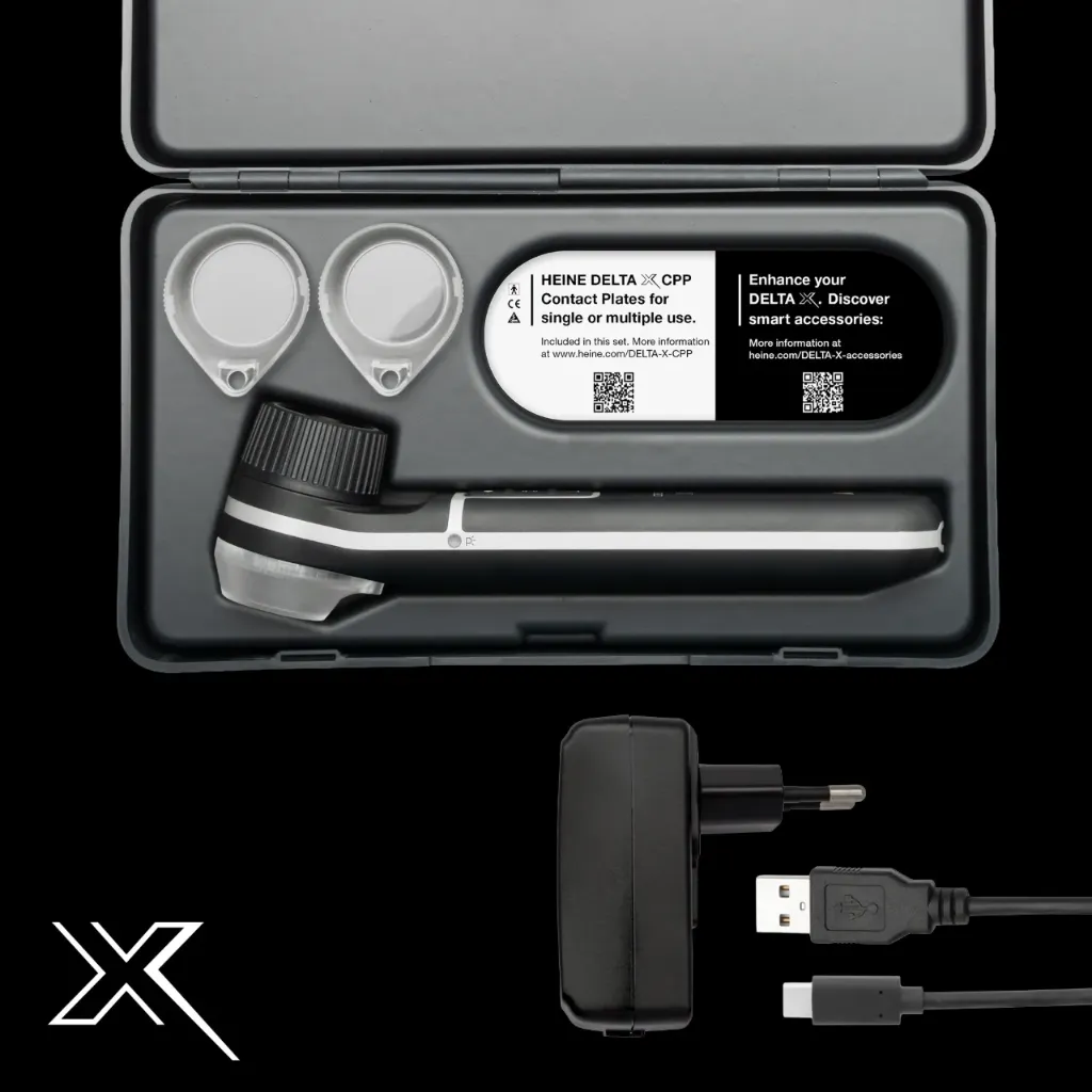
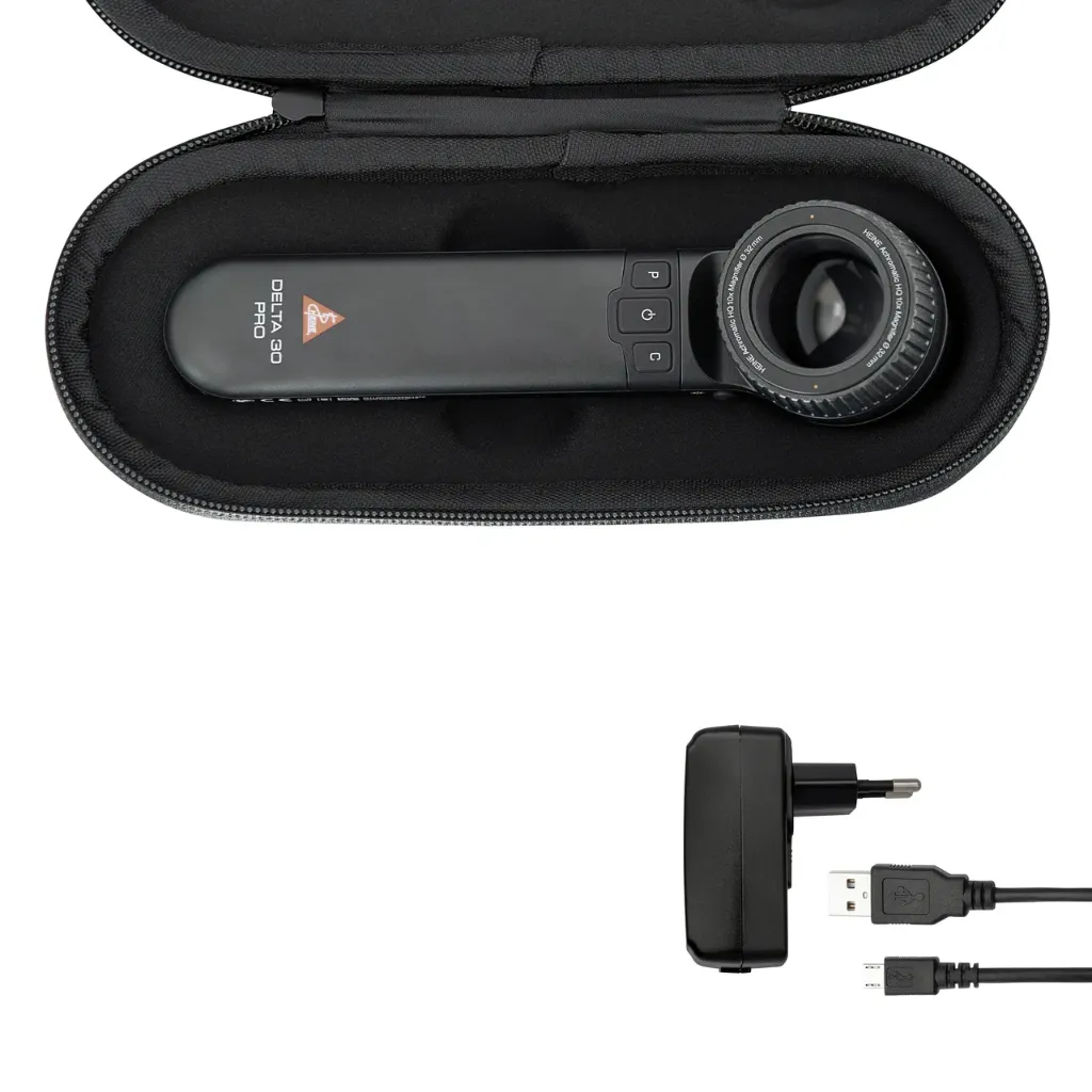
Photo: No photo Smartphone (optional) Camera (optional) Built-in camera
Specifications: HEINE and Casio technical data sheets.
Model by model: who is it for?
HEINE mini 3000. The most affordable pocket dermatoscope in the HEINE range: non-polarized light only, used in contact with an immersion fluid, 14 mm field and battery handle. Without a polarized mode, it does not show shiny white structures, and it offers no photo documentation. View the HEINE mini 3000
HEINE DELTA X. The most compact and lightest in the HEINE range, with the essentials: polarized / non-polarized switching, a small head for difficult areas, single-use contact plates and a magnetic smartphone adapter. Its 14 mm field, however, is the smallest in this comparison. View the HEINE DELTA X
HEINE DELTAone. A pocket dermatoscope with a 21 mm field, a good compromise between size and viewing comfort, for the practice as well as for home visits. View the HEINE DELTAone
HEINE DELTA 20T. The classic: 10x magnification, immersion possible in addition to polarized / non-polarized switching, and a DSLR camera adapter. It runs on a HEINE handle. View the HEINE DELTA 20T
HEINE DELTA 30. The large 30 mm field and comfortable battery life, for daily use and total body examination. View the HEINE DELTA 30
HEINE DELTA 30 PRO. The DELTA 30 with a revised LED layout and a color temperature adjustable in 4 steps, to adapt the rendering to your habits or to the lesion. View the HEINE DELTA 30 PRO
HEINE DELTA 30 UV. The DELTA 30 series format with additional UV lighting at 365 nm, for those who want to bring UV dermoscopy into their practice. View the HEINE DELTA 30 UV
Casio DZ-S50. A large 40.5 mm lens and non-contact examination, with an optional lens that brings magnification to 9x, which Casio highlights for small lesions such as those of scabies. Used without contact or fluid, it is mainly its polarized mode that gives a usable image. View the Casio DZ-S50
Casio DZ-D100. A dermatology camera rather than a dermatoscope: image quality comparable to a DSLR or mirrorless camera, plus images that are reproducible from one visit to the next, which makes lesion follow-up and teledermatology easier. View the Casio DZ-D100
Four situations, four starting points
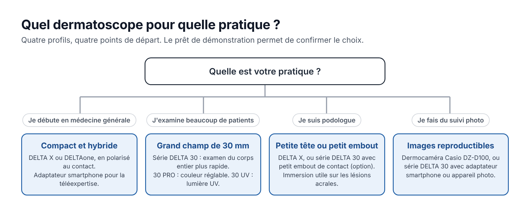
- You are starting dermoscopy in general practice. Start with training. As for equipment, a compact model with polarized / non-polarized switching (DELTA X or DELTAone) covers the essentials, with a smartphone adapter if you practice teledermatology.
- You examine many patients (dermatologist, trained general practitioner). The 30 mm field of the DELTA 30 series saves time during total body examination. The DELTA 30 PRO and DELTA 30 UV add color adjustment and UV respectively.
- You are a podiatrist. The areas to examine are often hard to reach: choose a small head (DELTA X) or a small contact plate, available as an option for the DELTA 30 series. Non-polarized dermoscopy with immersion is reported to perform better on acral lesions [9].
- Your priority is photo follow-up. A dedicated dermatology camera such as the DZ-D100, or a DELTA 30 series dermatoscope with an adapter, depending on your photo volume and the image management software you use.
Try before you choose
The best dermatoscope is the one you will use at every consultation. For dermatoscopes, a demo loan is available, as well as a remote presentation.
Also on our blog
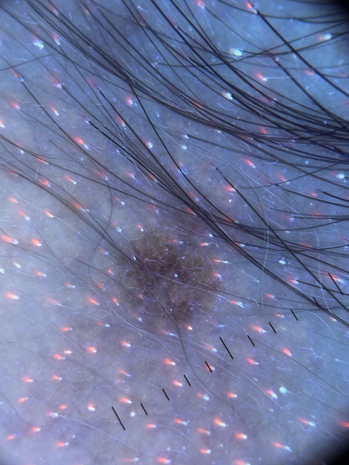
UV light in dermoscopy
What recent studies say about UV fluorescence and sub-UV reflectance.
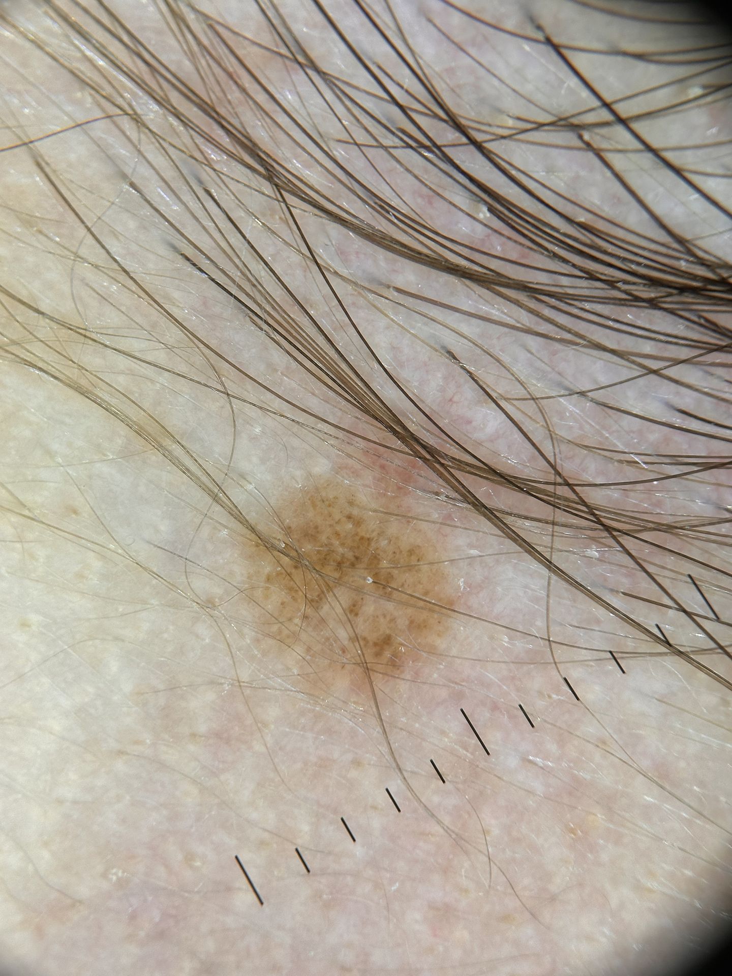
Polarized, non-polarized or immersion light
What each mode shows best, and what the studies say.
Sources
- Dinnes J, Deeks JJ, Chuchu N, et al. Dermoscopy, with and without visual inspection, for diagnosing melanoma in adults. Cochrane Database Syst Rev. 2018;12:CD011902. doi:10.1002/14651858.CD011902.pub2
- Vestergaard ME, Macaskill P, Holt PE, Menzies SW. Dermoscopy compared with naked eye examination for the diagnosis of primary melanoma: a meta-analysis of studies performed in a clinical setting. Br J Dermatol. 2008;159(3):669-676. doi:10.1111/j.1365-2133.2008.08713.x
- Argenziano G, Puig S, Zalaudek I, et al. Dermoscopy improves accuracy of primary care physicians to triage lesions suggestive of skin cancer. J Clin Oncol. 2006;24(12):1877-1882. doi:10.1200/JCO.2005.05.0864
- Benvenuto-Andrade C, Dusza SW, Agero AL, et al. Differences between polarized light dermoscopy and immersion contact dermoscopy for the evaluation of skin lesions. Arch Dermatol. 2007;143(3):329-338. doi:10.1001/archderm.143.3.329
- Dermoscopedia. Differences between polarized and non polarized dermoscopy. https://dermoscopedia.org/Differences_between_polarized_and_non_polarized_dermoscopy
- Williams NM, Rojas KD, Reynolds JM, et al. Assessment of diagnostic accuracy of dermoscopic structures and patterns used in melanoma detection: a systematic review and meta-analysis. JAMA Dermatol. 2021;157(9):1078-1088. doi:10.1001/jamadermatol.2021.2845
- Whybrew C, Pietkiewicz P, Kohut I, Chia JC, Akay BN, Rosendahl C. Not all polarized-light dermatoscopes may display diagnostically critical polarizing-specific features. Dermatol Pract Concept. 2022;12(4):e2022250. doi:10.5826/dpc.1204a250
- HEINE Optotechnik. HEINE DELTA 30 PRO, product brochure (EN). https://www.heine.com/fileadmin/data/Documents/HEINE-DELTA-30-PRO-brochure-EN.pdf
- Thompson H, Oakley A. A comparison of polarised and nonpolarised dermoscopy. DermNet, 2020. https://pro.dermnetnz.org/topics/a-comparison-of-polarised-and-nonpolarised-dermoscopy